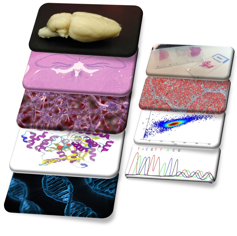
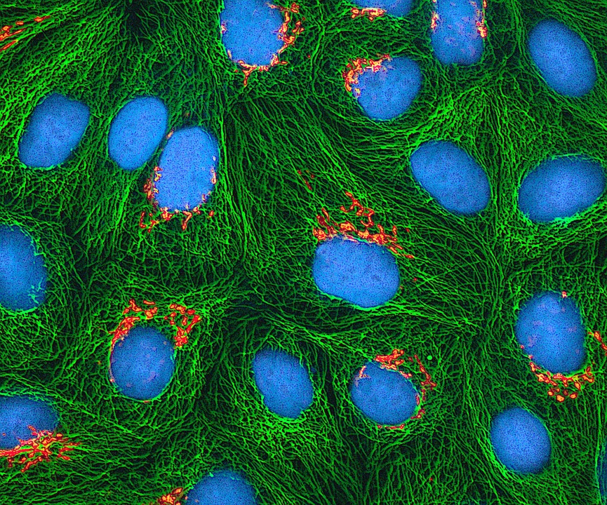
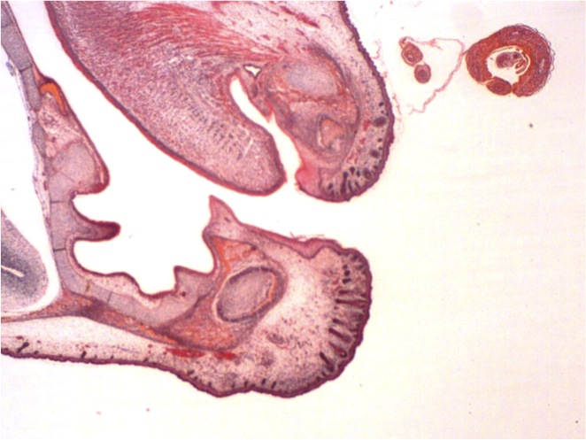
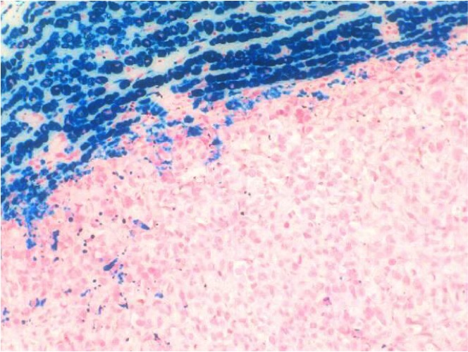
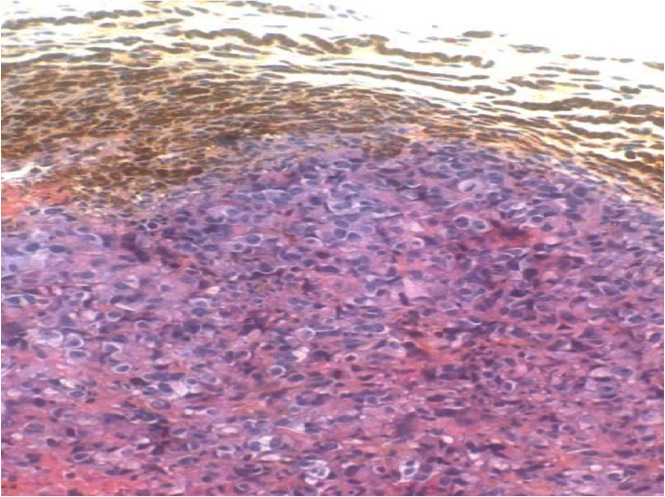
Our pathologists provide pathology report and scoring based on your own evaluation criteria. For each research field (Oncology, infectious disease, neuroscience … etc.), or matrix (animal, cells culture or 3D model… etc.) our pathologists will design a full analytical process including image analysis on visiopharm to ensure reliable and comprehensive results.
Our pathologists work with you for a global integration of histology, IHC, ISH, clinical pathology, molecular biology, and biochemistry results.
Our strength is our ability to complement standard histopathology evaluation with image analysis. Quantification by image analysis is the most effective way to compare cells population or protein expression in your samples.
All our image analysis tools are developed by the Dr Virgile Richard.
The morphological description of the standard staining lesions can be frequently complemented by quantitative, semi-quantitative or morphometric approaches. We are working on Image Pro Premier software packages and Visiopharm, this approach can complement a standard analysis and allows you to have numerical values in accordance with the qualitative examination of the pathologist. All the image analysis process is done by the Dr Virgile Richard.
Whole slide image analysis is a complex process and implies knowledge of physiopathology mechanisms involved in the model used. That’s why image analysis at Biodoxis is not only usage of informatics tools. All relevant morphological or pathological aspects associated with a model are taken in account to give reliable and accurate data. Image analysis is carried out with the same systematic multi-magnification approach used by a pathologist during his histopathological examination.
It is now possible to measure, localize and characterize any tissue changes (fibrosis, angiogenesis, apoptosis, hypertrophy/hyperplasia, inflammation or else), specific protein expression, and specific cell type density in a whole tissue context. This approach corresponds to the commonly described object-oriented image analysis.
Sciempath Labo can work with you to develop a personalized image analysis approach to fulfill your expectations.
A classic histopathological analysis will give answers on which compound have a beneficial effect on your tissue. Image analysis performed by a pathologist can give you a numeric value to compare the effectiveness of several treatments on different biological parameters (cell phenotyping, protein expression, or morphological changes).
The image analysis coupled with histopathology will help extracting the most out of the same experimental material. Our pathologists are available to advise you on the use of image analysis tools.